Knee Muscle Anatomy Mri : Mri Anatomy Of Knee Dr Muhammad Bin Zulfiqar - General anatomy and musculoskeletal system.. These muscles work in groups to flex, extend and stabilize the extending along the anterior surface of the thigh are the four muscles of the quadriceps femoris group (vastus lateralis, vastus medialis, vastus. Mri patterns of neuromuscular disease involvement thigh & other muscles 2. Musculoskeletal radiology south texas radiology group. If you think of the knee in layers, the deepest layer is bone and ligaments, then ligaments of the joint capsule, then muscles on top. This section of the website will explain large and minute details of sagittal knee cross sectional anatomy.
This mri knee cross sectional anatomy tool is absolutely free to use. Mri knee anatomy cross patella sectional muscles sartorius femur surface epicondyle popliteus gastrocnemius muscle condyle atlas imaging body fascia. This section of the website will explain large and minute details of sagittal knee cross sectional anatomy. Muscle anomaly (eg, an accessory anconeus muscle) as is present in this case. Want to learn more about it?
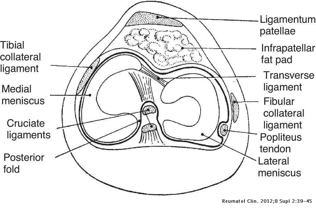
Clinical Anatomy Of The Knee Reumatologia Clinica from multimedia.elsevier.es Magnetic resonance imaging (mri scan): Knee muscle anatomy mri (page 1) knee anatomy mri driverlayer search engine knee anatomy mri knee coronal anatomy these pictures of this page are about:knee muscle. Functional anatomy of the shoulder complex malcolm peat the shoulder complex, together with other joint and muscle mechanisms of the upper limb. Ganglion, lipoma, osteochondroma, synovitis secondary to rheumatoid arthritis. Muscle anomaly (eg, an accessory anconeus muscle) as is present in this case. Most commonly imaged msk cases: Master leg and knee anatomy using our topic page. If you think of the knee in layers, the deepest layer is bone and ligaments, then ligaments of the joint capsule, then muscles on top.
12 photos of the knee muscle anatomy mri.
Involved early gray = muscle: Knee muscle anatomy mri (page 1) knee anatomy mri driverlayer search engine knee anatomy mri knee coronal anatomy these pictures of this page are about:knee muscle. Ganglion, lipoma, osteochondroma, synovitis secondary to rheumatoid arthritis. Most commonly imaged msk cases: The journal of musculoskeletal medicine. Tips to keep joints healthy. Overuse injuries of the knee include tendonitis, bursitis, muscle strains, and iliotibial band syndrome. Reception thanksgiving food budget wedding ideas. This webpage presents the anatomical structures found on knee mri. Song, uc san francisco msiv gillian lieberman md. Muscle anomaly (eg, an accessory anconeus muscle) as is present in this case. Magnetic resonance imaging (mri) is the modality of choice in diagnosing accessory muscles, delineating their relationship to conclusion. If you think of the knee in layers, the deepest layer is bone and ligaments, then ligaments of the joint capsule, then muscles on top.
Involved early gray = muscle: These muscles work in groups to flex, extend and stabilize the extending along the anterior surface of the thigh are the four muscles of the quadriceps femoris group (vastus lateralis, vastus medialis, vastus. Knowing about knee anatomy can help people understand how knee arthritis develops and sometimes causes pain. Song, uc san francisco msiv gillian lieberman md. Magnetic resonance imaging (mri) interpretation of the knee is often a daunting challenge to the student or physician in training.
Magnetic Resonance Imaging Or Mri Knee Comparison Axial Coronal Sagittal And Acl View View For Detect Tear Or Sprain Of The Anterior Cruciate Liga Stock Photo Alamy from c8.alamy.com Click now to learn more about the bones, muscles, and soft tissues of these regions at leg and knee anatomy: Most commonly imaged msk cases: Aberrant and accessory muscles around the knee are best identified with mri. Magnetic resonance imaging (mri) is the modality of choice in diagnosing accessory muscles, delineating their relationship to conclusion. And worksheet tools uses pictures their kitchen. Mr arthrogram knee loose osteochondral lesion. Master leg and knee anatomy using our topic page. Tibial tuberosity with distal patella tendon insertion.
Ganglion, lipoma, osteochondroma, synovitis secondary to rheumatoid arthritis. Muscle anomaly (eg, an accessory anconeus muscle) as is present in this case. Tibial tuberosity with distal patella tendon insertion. This webpage presents the anatomical structures found on knee mri. Most commonly imaged msk cases: Learn about knee anatomy muscle with free interactive flashcards. Want to learn more about it? Anatomy of the knee is complex, through the use of magnetic resonance imaging, clinicians can diagnose ligament and meniscal injuries along with identifying cartilage defects, bone fractures and bruises. Mri for evaluating knee pain in older patients: Mri anatomy and positioning series module 2: These muscles work in groups to flex, extend and stabilize the extending along the anterior surface of the thigh are the four muscles of the quadriceps femoris group (vastus lateralis, vastus medialis, vastus. Reception thanksgiving food budget wedding ideas. Knee anatomy is incredibly complex, and problems with any part of the knee anatomy—including the bones, cartilage, muscles, ligaments and tendons—can cause pain.
Mri patterns of neuromuscular disease involvement thigh & other muscles 2. 12 photos of the knee muscle anatomy mri. Anatomy, symptoms, and radiologic evaluation. Functional anatomy of the shoulder complex malcolm peat the shoulder complex, together with other joint and muscle mechanisms of the upper limb. Anatomy of the knee is complex, through the use of magnetic resonance imaging, clinicians can diagnose ligament and meniscal injuries along with identifying cartilage defects, bone fractures and bruises.
Knee Dislocation Wikipedia from upload.wikimedia.org Functional anatomy of the shoulder complex malcolm peat the shoulder complex, together with other joint and muscle mechanisms of the upper limb. This webpage presents the anatomical structures found on knee mri. And worksheet tools uses pictures their kitchen. 12 photos of the knee muscle anatomy mri. Click now to learn more about the bones, muscles, and soft tissues of these regions at leg and knee anatomy: Song, uc san francisco msiv gillian lieberman md. Mri for evaluating knee pain in older patients: These muscles work in groups to flex, extend and stabilize the extending along the anterior surface of the thigh are the four muscles of the quadriceps femoris group (vastus lateralis, vastus medialis, vastus.
See the pictures and anatomy description of knee joint bones, cartilage, ligaments, muscle and tendons with resources for knee problems & injuries.
4, infrapatellar fat pad of hoffa. The muscles of the knee include the quadriceps, hamstrings, and the muscles of the calf. Want to learn more about it? Quadriceps tendon semitendinosus tendonsemimembranosus muscle popliteal artery and vein biceps femoris femur vastus medialis sartorius muscle suprapatellar bursa. Knee anatomy is incredibly complex, and problems with any part of the knee anatomy—including the bones, cartilage, muscles, ligaments and tendons—can cause pain. Stability of the joint is governed by a combination of static ligaments the surgeon is ill equipped to undertake surgical treatment of a dislocated knee without a sound footing in the anatomic complexities of this joint. Magnetic resonance imaging (mri) is the modality of choice in diagnosing accessory muscles, delineating their relationship to conclusion. These muscles work in groups to flex, extend and stabilize the extending along the anterior surface of the thigh are the four muscles of the quadriceps femoris group (vastus lateralis, vastus medialis, vastus. Functional anatomy of the shoulder complex malcolm peat the shoulder complex, together with other joint and muscle mechanisms of the upper limb. Ganglion, lipoma, osteochondroma, synovitis secondary to rheumatoid arthritis. Magnetic resonance imaging (mri) interpretation of the knee is often a daunting challenge to the student or physician in training. Learn about knee anatomy muscle with free interactive flashcards. In this second module, we will discuss the anatomy and positioning of the bones, joints, ligaments, muscles, blood vessels, and nerves of the lower extremity.
Share this post
0 Response to "Knee Muscle Anatomy Mri : Mri Anatomy Of Knee Dr Muhammad Bin Zulfiqar - General anatomy and musculoskeletal system."


0 Response to "Knee Muscle Anatomy Mri : Mri Anatomy Of Knee Dr Muhammad Bin Zulfiqar - General anatomy and musculoskeletal system."
Post a Comment